Complications: What You Need to Know and How to Prevent Them Effectively
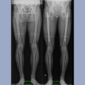
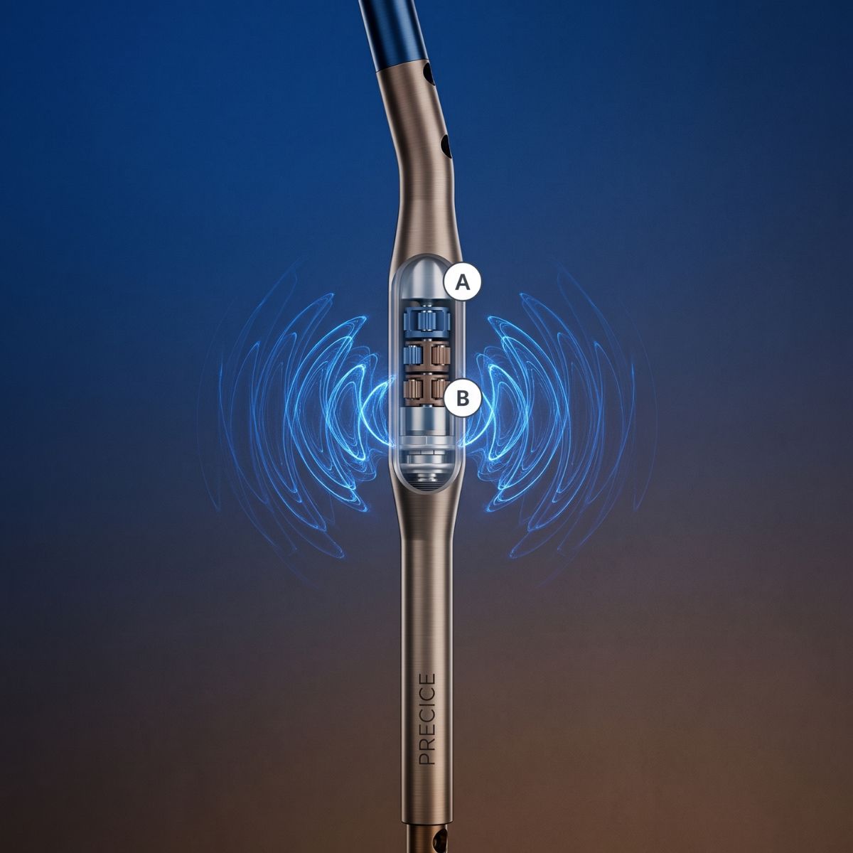
Every procedure carries some risk, but complications are extremely rare, and thanks to the use of modern internal nails (such as PRECICE), the incidence of complications is significantly lower than with external methods such as LON.
Even in the face of unforeseen challenges, our team has extensive experience in effectively and safely managing any complications to the highest global standards.
We are committed to full transparency, which is why we have provided a detailed list of potential complications below.
Like any surgical procedure, limb lengthening carries certain risks; however, it must be strongly emphasized that complications are extremely rare. Their incidence is closely related to the chosen treatment method. Historically, when using traditional external fixators and the LON method (lengthening over an intramedullary nail), the risk of certain problems—such as frequent infections around the implants (so-called wires)—was noticeably higher.
However, the situation is completely different when using modern, fully internal magnetic nails, such as PRECICE. Because the entire device is located inside the bone, the rate of complications drops dramatically—for example, the risk of infection with the PRECICE system is very low, at less than 1%. Furthermore, intramedullary nails provide excellent mechanical control over the rate of lengthening, which minimizes the risk of dangerous stresses, muscle contractures, or nerve damage.
Despite the highest standards of prevention, medicine can never offer a 100% guarantee that a patient’s body will not react in an unforeseen way (e.g., by healing too slowly or too quickly). And it is precisely in such moments that our experience is crucial. As we emphasize: for a surgeon, it is not only extremely important to perform the surgery correctly, but above all to manage all potential complications efficiently and effectively.
We are prepared for any scenario, no matter how rare. Whether it requires adjusting the rate of limb lengthening, specialized medication, the use of bone growth stimulators, or a minor corrective procedure—we can handle any complication at the highest international standard, ensuring patients’ complete safety and the ultimate success of the entire treatment.
DVT can occur after any orthopedic surgery or fracture. Fortunately, we have a very low rate of this complication (1%). Prevention is key. We prescribe children’s aspirin (81 mg) twice daily after surgery, both in the hospital and on an outpatient basis, until the distraction phase is complete. Although we have seen very few cases of DVT, none have resulted in pulmonary embolism (PE). PE occurs when a blood clot breaks loose and travels to the lungs. This can cause shortness of breath, chest pain, and even death. That is why we take great care to protect our patients from this. Taking oral contraceptives and smoking increase the risk of DVT. All our patients receive anticoagulant medication—either aspirin or Xarelto for high-risk patients.
To insert a Precice nail into the bone, the surgeon must drill into the bone in a process called bone reaming. Reaming leads to increased pressure within the bone (similar to pushing a plunger into a syringe), which displaces the fat located in the bone marrow. Some of this fat is forced into the bloodstream. When excess fat reaches the lungs and becomes trapped there, this is called a fat embolism. Small amounts of fat are well tolerated by the lungs and do not cause any symptoms.
Larger amounts can lead to shortness of breath and lower blood oxygen levels; in some cases, they can cause coughing or even loss of consciousness. In most cases, this is treated by administering oxygen through a nasal cannula for several days. In more severe cases, an oxygen mask is used instead. In very rare and severe cases of fat embolism syndrome (FES), the patient may need to be transferred to the ICU.
Patients undergoing limb-lengthening procedures require not only rapid diagnosis and treatment if symptoms of fat embolism syndrome (FES) occur, but above all, the implementation of effective preventive strategies to minimize the risk of this complication. Over the years, the Paley European Institute has developed a multi-step preventive protocol that has reduced the incidence of FES to less than 0.1%. In total, only two cases of fat embolism have been reported over the past 7 years.
A key element of prevention is the use of specialized sharp reamers, which allow for decompression of the intramedullary canal while reaming it. This reduces intramedullary pressure, and the fatty-medullary material—which might otherwise enter the venous circulation—is evacuated through the reamer’s insertion site.
In addition, Dr. Dror Paley developed a technique for creating ventilation holes at the level of the osteotomy, which provide an additional route for decompressing the bone canal during reaming and intramedullary nail implantation.
At our center, this protocol has been expanded to include an additional proprietary modification: the creation of an extra ventilation hole in the distal femur, combined with the use of suction and controlled negative pressure. This technique allows for the active removal of excess fat, bone marrow, and tissue debris that accumulate during drilling, which further reduces intramedullary pressure and minimizes the risk of embolization.
Another significant advantage of this method is the reduction in heat generated during reaming, which may limit thermal damage to the bone and intraosseous vessels. This may potentially have a beneficial effect on the maintenance of intraosseous perfusion and the quality of the bone regenerate during the subsequent distraction process.
All of the techniques mentioned have proven to be exceptionally effective. Importantly, since the implementation of an additional ventilation port with active suction at the Paley Lengthening Center, not a single case of fat embolism syndrome has been reported.
Slow bone healing or a lack thereof can occur with any limb-lengthening surgery. The best treatment is prevention. We begin by identifying factors that may slow healing even before surgery: osteoporosis, rickets, menopause, low vitamin D levels, smoking/tobacco use (including secondhand smoke), and the use of anti-inflammatory, anticonvulsant, and other medications. We also recommend supplements that help bones heal faster.
Since 2011, when we began using the Precice nail, not a single patient has required surgery due to delayed or failed union. All patients were successfully treated with non-surgical methods. If delayed healing occurs despite the above steps, we begin using the “accordion” technique. Using the ERC device, the bone is compressed by one mm per day and stretched by one mm per day. This cycle is repeated several times a day, which stimulates bone healing and helps avoid surgery.
If surgery is necessary, we know how to promote bone healing through surgical means. We gained this experience using older implants that lacked rate control (e.g., ISKD), where nonunion was not uncommon. As a result, we have become highly skilled in bone and bone marrow transplants. Although we hope we will never need to use these techniques with Precice nails, we are highly experienced in managing this complication should the need arise. It is crucial for a surgeon not only to know how to perform the surgery but also how to manage all potential complications.
Nerve damage can occur with any limb-lengthening surgery, but it is quite rare if the distraction rate does not exceed 1 mm per day and the extent of lengthening is limited. Controlling the distraction rate is the most important factor in preventing nerve damage. It is important to recognize neurological symptoms.
In such cases, lengthening should be stopped or slowed down. If any motor symptoms (muscle weakness or paralysis) occur, nerve decompression surgery should be performed as soon as possible. This is a minor outpatient procedure. In most cases, the problems involve the peroneal nerve. It is important for the surgeon to know how to decompress this nerve to prevent foot drop. Delaying decompression can lead to permanent foot drop. Thanks to Precice nails, which allow for excellent control of the advancement rate, nerve damage is very rare. Nerve irritation can also be treated with medication.
A specific, minor nerve injury involves damage to the small branch that innervates the extensor hallucis longus muscle. This nerve is at risk during fibular osteotomy, which we perform as part of tibial lengthening. This nerve injury leads to drop foot of the big toe, which causes almost no noticeable problems. Although this complication is very rare, if it occurs, there is no treatment other than waiting for the nerve to regenerate on its own, which usually happens.
Muscles typically become tense when they are stretched. A muscle contracture occurs when a muscle tightens so much that it prevents the joint from moving through its full range of motion. Physical therapy (PT) is essential for preventing muscle contractures.
The patient should perform daily stretching exercises for the muscles and joints at risk (e.g., the knee joint and quadriceps muscles during femur lengthening, and the ankle joint and Achilles tendon during tibia lengthening). In addition to formal physical therapy, the patient should stretch independently at home several times a day. Physical therapy is crucial to the lengthening process. However, it is expensive. I do not consider performing lengthening if the patient is not willing to undergo rehabilitation. This is not an option for reducing costs. The controlled rate of lengthening provided by the Precice system reduces the risk of contractures and muscle spasms; however, Precice does not eliminate the need for physical therapy. Maintaining range of motion and preventing contractures during lengthening shortens the rehabilitation time needed to return to normal functioning after the process is complete.
A permanent knee or ankle contracture can lead to disability and the need for prolonged physical therapy and the associated costs. If the contracture persists despite additional rehabilitation, further surgery to lengthen muscles, tendons, and fascia may be required. We strive to anticipate this and proactively lengthen certain soft tissue structures to prevent contractures. If this is done during the initial surgery, the additional cost is minimal. If soft tissue lengthening surgery is required at a later date, the cost is significantly higher due to hospital fees.
While lengthening certain muscles or tendons has no adverse effects, some tendon lengthening procedures can lead to permanent weakness and should be avoided. Lengthening this structure in an adult often leads to permanent weakness in the push-off force, which affects running and jumping. Some surgeons freely lengthen this structure during tibial lengthening to achieve a greater increase in length. We avoid this procedure because the patient would never regain their previous ability to run and jump. For this reason, we typically limit tibial lengthening to 5 cm, which avoids the need to lengthen this muscle/tendon.
When lengthening the tibia, the fibula must also be lengthened. The implanted lengthening device only lengthens and stabilizes the tibia. The fibula must be attached to the tibia so that they lengthen together. If the fibula is not attached or is attached improperly, it will not lengthen as much as the tibia, leading to serious consequences, including subluxation and inflammation of the ankle joint, as well as flexion contracture of the knee.
The stabilization method is crucial. Many surgeons fix only the distal end of the fibula to the tibia. This can lead to premature union of the fibula, which then pulls downward and shifts relative to the tibia at its proximal end. It is important to fix the fibula at both ends. Most surgeons avoid this because it is technically difficult to perform.
For all of our patients, we secure the fibula at both ends. During implant-based lengthening, the fibula must be secured to the tibia with screws—one screw at the upper end and one at the lower end. The angle, level, position, diameter, and type of screw—all of these are important. A common mistake is placing the screw horizontally between the two bones. This is not a strong enough connection to prevent the fibula from separating from the tibia at the ankle. This is a very subtle issue, and even a difference of just a few millimeters in the length of the fibula at the ankle can lead to short- and/or long-term consequences for the patient. Removing a segment of the fibula to prevent it from separating is another common method that should be avoided. This leads to nonunion of the fibula, which can later result in a stress fracture of the tibia.
Furthermore, this usually does not prevent the fibula from drifting away from the tibia. Therefore, complications involving the fibula have nothing to do with the type of lengthening implant used, but rather with the method the surgeon chooses to stabilize the fibula relative to the tibia and the way the fibula is cut. Healing of the fibula can range from premature union to complete nonunion. The latter often does not require treatment, provided that the tibia has healed soundly. The former requires a repeat osteotomy of the fibula.
Axial deviation or secondary deformity associated with lengthening is not an uncommon problem. It can usually be prevented. Varus deformity is more likely to occur when using a trochanteric nail. For this reason, I prefer to use a nail inserted through the piriformis foramen. In cases of proximal femoral lengthening, if the osteotomy is performed at a site where the bone is tight around the nail, the likelihood of varus angulation is low. If a more proximal femoral osteotomy is necessary, a medial blocking screw can prevent varus deviation.
Axial deviation is a more common problem in the tibia. The tibia tends to elongate in the direction of valgus and procurvatum. To prevent this, the nail should be inserted at the correct starting point, which is located very high up and as far posteriorly on the tibia as possible on the lateral view, and centered on the knee in the frontal plane. If there is a gap between the nail and the lateral cortex above the osteotomy, a locking screw should be inserted into it. If there is a space between the nail and the posterior cortex, another locking screw is placed there. I designed the nail to resist axial deviations, also providing a third proximal locking screw. If all these precautions are followed, no flexion or valgus deformity develops during lengthening.
There is a small risk that the lengthened bone may fracture after the device is removed. If this happens, another surgery or the use of a cast or brace may be necessary. For this reason, it is extremely important to follow your doctor’s recommendations regarding physical activity.
This is the most common complication associated with treatment using an external fixator. It manifests as tenderness, redness, a sensation of warmth, swelling, pain, or exudate at the site where the wires were inserted. It requires prompt (within 24 hours) initiation of oral antibiotic therapy. If left untreated, the infection can spread to deeper tissues and even reach the bone. A specific risk factor for the LON (lengthening over an intramedullary nail) technique is that an infection originating around the implants can spread to the implanted nail and the entire medullary canal.
Photo gallery